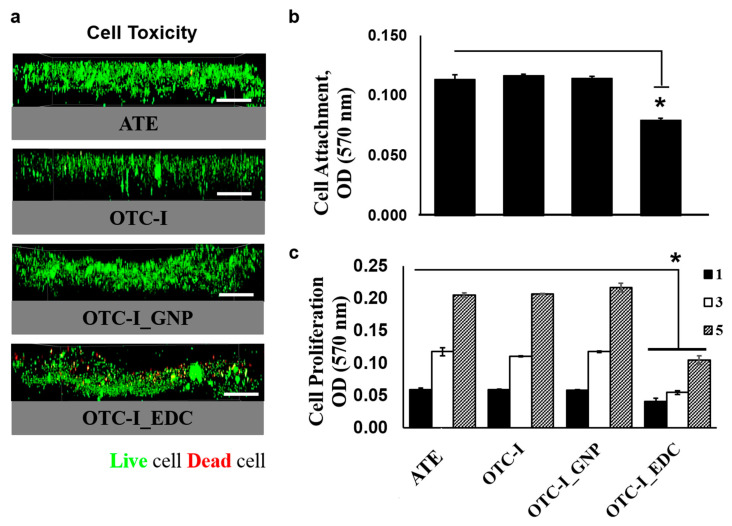
Figure 4.
Immunochemical (a) and the HDF attachment (b) and growth pattern (c) of skin cells seeded on fabricated collagen bioscaffold at day 1, 3 and 5 (bar = 100 μm). The skin cells viability well-distributed on each collagen bioscaffold except the group crosslinked with carbodiimide (EDC). The skin cells growth gradually increased following time for all treatment groups and significantly lower in the group crosslinked with carbodiimide (EDC). * represents statistically significant difference (p < 0.05).

