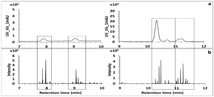
Figure 7.
Screening of MAAs in the model alga Gymnogongrus devoniensis. UV Trace of MAAs recorded at (300–360) nm (a). XIC Trace MS2 of the set of eight fragment ions by ddMS2/MS3 untargeted analysis (b). All the peaks observed in the XIC trace MS2 indicate all the retention times for which MAAs were detected in the extract.

