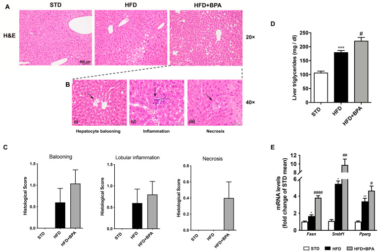
Figure 2.
BPA exposure worsens hepatic steatosis and inflammation in HFD-fed mice. (A) H&E staining was performed on the liver from all groups of animals (magnification 20×) (n = 4 each group). (B) (i) The appearance of the ballooning degeneration, (ii) the inflammatory and (iii) necrotic lesions in the liver from HFD + BPA, and (C) the histological score for all three groups were shown. (D) Hepatic triglycerides and mRNA expression of (E) Fasn, Srebf1, and Pparg were determined. Data are presented as means ± SEM of all animals (n = 6 each group) (* p < 0.05 vs. STD, ** p < 0.01, *** p < 0.001, # p < 0.05, ## p < 0.01, and #### p < 0.0001 vs. HFD).

