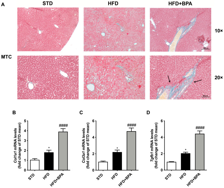
Figure 5.
BPA exposure promotes liver fibrosis progression in HFD-fed mice. (A) Mallory trichrome (MTC) staining was assessed to determine the fibrotic lesions induced by BPA on the liver from HFD mice (n = 4 each group). Gene expression of pro-fibrotic (B–D) Col1a1, Col3a1, and Tgfb was determined by real-time PCR analysis. Data are presented as means ± SEM of all animals (n = 6 each group) (* p < 0.05 vs. STD; #### p < 0.0001 vs. HFD).

