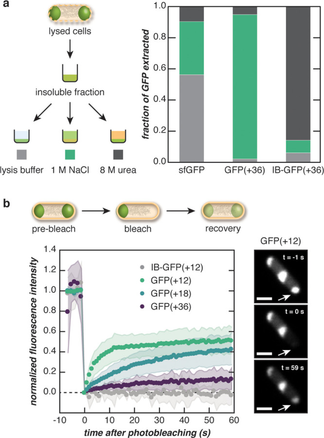
Figure 3.

Fluorescent puncta behave as complex coacervates. (a) The dense, insoluble fraction of lysed E. coli cells expressing engineered GFPs was solubilized in a range of buffers to distinguish the behavior of supercationic GFP(+36) from an inclusion body (IB)-forming variant. The insoluble fraction was treated with lysis buffer, 1 M NaCl, or 8 M urea, and the fraction of GFP (sfGFP, GFP(+36), or IB-GFP(+36)) solubilized with each treatment was determined by SDS-PAGE analysis. (b) One pole of an E. coli cell was bleached, and the fluorescence recovery was monitored over time (left). The panels show the fluorescence of a representative cell expressing GFP(+12) at different time points during FRAP (right). Supercharged GFP droplets were dynamic relative to IB-forming GFPs and became less dynamic with increasing protein charge. Scale bar, 1 μm.
