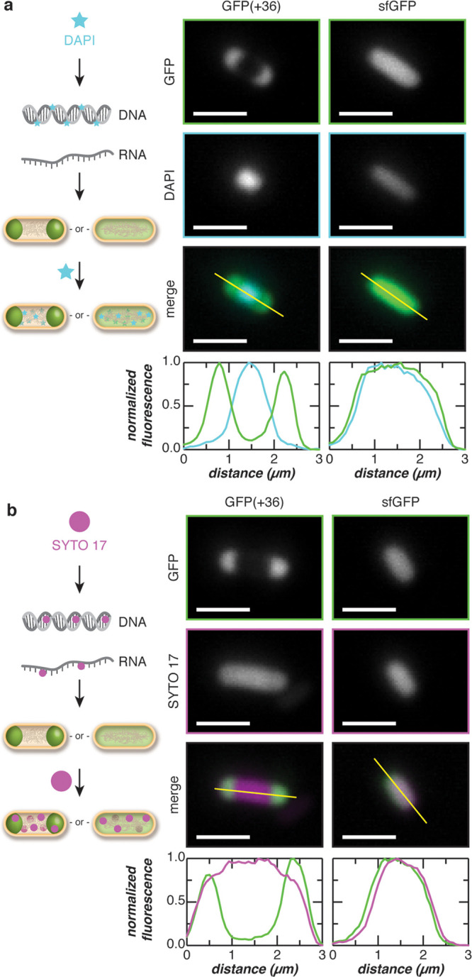
Figure 4.

Colocalization of endogenous nucleic acids with supercationic GFP condensates. (a) The colocalization of DNA with the GFP(+36) condensates was evaluated by staining cells with DAPI (a DNA-specific dye). Microscopy images depict cells expressing GFP(+36) or sfGFP and stained with DAPI. Intensity line-cuts demonstrate exclusion of DNA from the GFP(+36) condensates. (b) The colocalization of RNA with the GFP(+36) condensates was evaluated by staining cells with SYTO 17, which binds both DNA and RNA. Microscopy image of cells expressing GFP(+36) or sfGFP and stained with SYTO 17 are shown along with intensity line-cuts demonstrating colocalization of RNA and GFPs. Scale bars, 2 μm.
