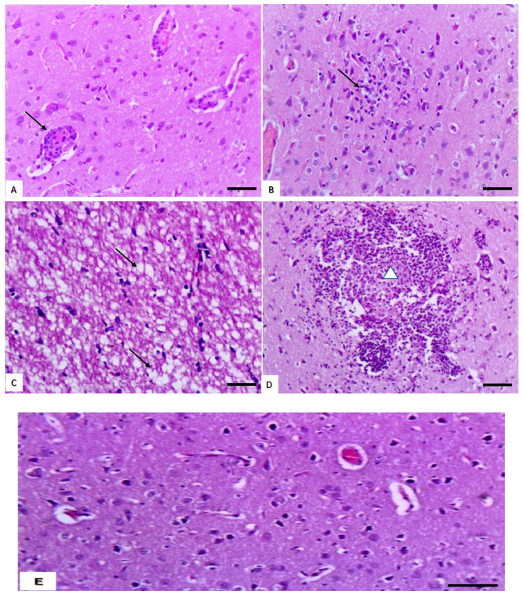
Figure 3.
Effect of cymoxanil on the brain. (A) Brain of rats treated with medium dose showing perivascular cuff (arrows) (H&E, bar = 50 µm). (B) Brain of rats treated with medium dose showing slight gliosis (arrow) (H&E, bar = 50 µm). (C) Brain of rats treated with high dose showing cerebral spongiosis (arrow) (H&E, bar = 50 µm). (D) Brain of rats treated with high dose showing cerebral malacia (arrow) (H&E, bar = 50 µm). (E) Brain of control showing intact neuronal cells and blood vessels surrounded with clear perivascular space in the neuropil of cerebrum (H&E, bar = 50 µm).

