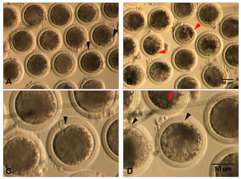
Figure 2.
Denuded MII bovine oocytes after 24 h of IVM. (A,C) oocytes showing a homogeneous dark cytoplasm. Black arrows depict the first polar body; (B,D) oocytes showing a heterogeneous pale and punctuated cytoplasm. Black arrows indicate the first polar body, while red arrows depict dark areas of intense lipid accumulation (cytoplasmic granulations).

