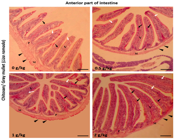
Figure 1.
Histomicrograph showing the histological structures of the anterior part of Grey Mullet intestines in the control group and chitosan-fed groups (0.5, 1, and 2 g/kg). The intestine shows normal histological structures of the intestinal wall and intestinal villi in all groups. The intestinal wall was formed of tunica mucosa of normally arranged enterocyte (white arrow) and goblet cells (black arrow), propria submucosa (P), tunica muscularis (M), and tunica serosa (black arrowhead). The villous height, width, and the number of goblet cells increased gradually in a dose-dependent manner with chitosan. Stain periodic acid-Schiff (PAS). Bar = 200 µm.

