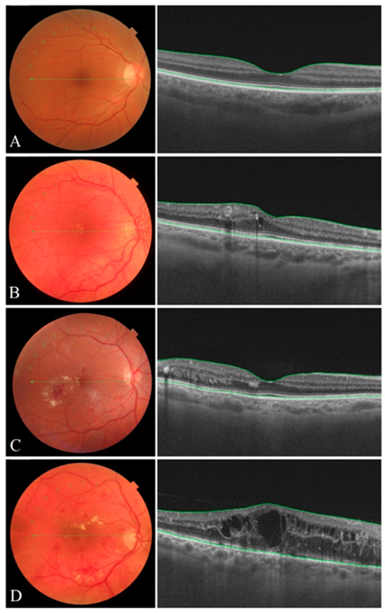
Figure 1.
Color eye fundus images of the human retina (left) and high–quality SD OCT images (right) of the macula representing various stages of DR severity: (A) mild DR, (B) moderate DR, (C) severe DR, (D) proliferative DR. Mild DR is characterized by microaneurysms, moderate DR is distinguished by more microaneurysms but less than seen in severe DR. Signs of severe DR include the following: dot-and–blot-hemorrhages, venous beading, cotton wool spots, exudates and intraretinal microvascular abnormalities (IRMA). A clinical sign of proliferative DR is neovascularization, which can lead to macular edema.

