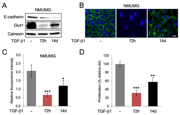
Figure 3.
Prolonged TGF-β1 exposure is associated with more pronounced EMT, re-expression of Glut1, and increased glucose uptake and cell proliferation. (A) Western blot analysis of the expression of E-cadherin and Glut1 in NMuMG cells after short-term (72 h) and long-term (14 days) exposure to TGF-β1. Calnexin was used as a loading control. (B) Immunofluorescence staining of Glut1 in NMuMG cells after short-term (72 h) and long-term (14 days) TGF-β1 exposure. DAPI staining was used to visualize cell nuclei. Scale bar = 10 μm. (C,D) Bar graphs showing the effects of short-term (72 h) and long-term (14 days) exposure of NMuMG cells to TGF-β1 on the uptake of 2-NBDG (C) and cell proliferation (D). * p < 0.05; ** p < 0.01; *** p < 0.001.

