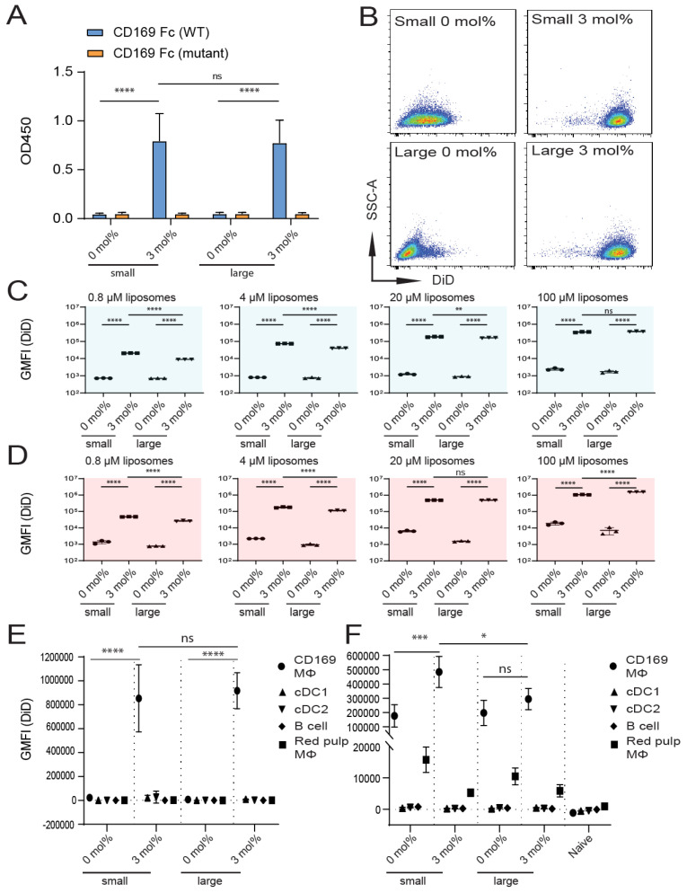
Figure 4.
Size of GM3-containing liposomes influences binding to CD169-expressing cells in vivo, but not in vitro. (A) Liposomes were coated overnight on an ELISA plate, and binding to WT or mutant CD169 Fc was quantified. Indicated is the average OD450 of 2 independent technical triplicates. (B–D) DiD-containing liposomes were incubated with TSn at 4 or 37 °C for 45 min. Representative dot plots following incubation at 37 °C (20 µM liposomes) (B). Indicated is the average GMFI ± SD of a technical triplicate after incubation at 4 °C (C) and at 37 °C (D) (representative of 4 independent experiments). (E) DiD-containing liposomes were incubated for 45 min at 37 °C with freshly isolated splenocytes from C57BL/6 WT mice. Subsequently, DiD fluorescence was quantified for distinct cell populations using flow cytometry. Indicated is the GMFI ± SD (n = 4) (representative of 2 independent experiments). (F) DiD-containing liposomes (22.5 nmol of phospholipid) were injected IV in mice, and after 2 h, splenic cell populations were analyzed for DiD fluorescence. Indicated is the average GMFI ± SD (n = 4) (F). * p < 0.05, ** p < 0.01, *** p < 0.005 and **** p < 0.0001, ns: no significance.

