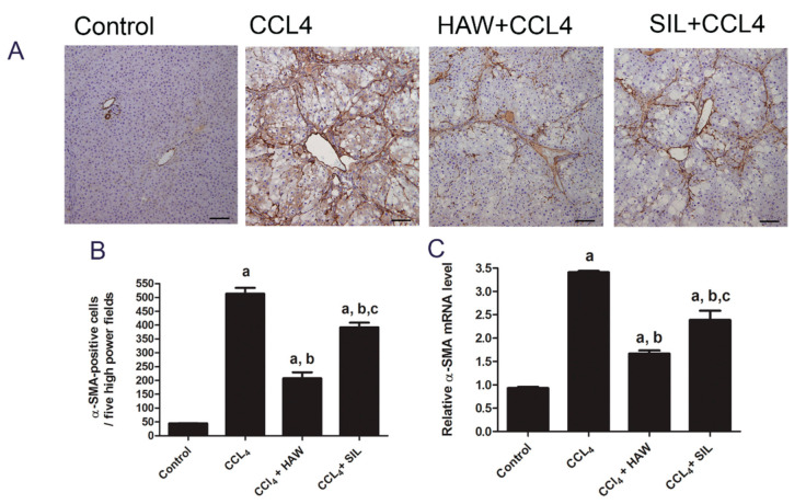
Figure 3.
Effects of HAW on the CCl4-induced hepatic stellate cells (HSC) activation in liver fibrosis model. HAW inhibited HSC activation in liver fibrosis. (A) representative images of immunohistochemically expression of α-SMA in the liver was determined by immunohistochemistry (brown area), 100× magnification, scale bar equals 100µm. In CCL4 section marked staining for alpha-smooth muscle actin (α-SMA) is found along with the fibrous septa. (B) the expression of α-SMA cells in each section was calculated by counting the number of brown staining, α-SMA-positive cells in five fields per section at 400× magnification, n = 6 per group). (C) the mRNA expression levels of α-SMA were detected by q-RTPCR. Data are presented as means ± SEM, n = 3 per group. a is p < 0.05 vs. control group, b is p < 0.05 vs. CCl4 group, and c is p < 0.05 vs. HAW group.

