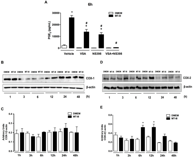
Figure 2.
MT-III activates COX-1 and COX-2 pathways for release of PGE2 by 3T3-L1 preadipocytes. (A) Cells were incubated with either valerylsalicylate (VSA) (10 µM), or NS-398 (10 µM), or both for 1 h, followed by incubation with MT-III (0.4 µM) for 6 h. PGE2 concentrations were quantified in culture supernatants by EIA commercial kit. (B–E) 3T3-L1 preadipocytes were incubated with MT-III (0.4 µM) or DMEM (control) for 1 up to 48 h. (B) Western blotting of COX-1 and β-actin (loading control) showing immunoreactive bands. (D) Western blotting of COX-2 and β-actin (loading control) showing immunoreactive bands. Densitometric analysis of immunoreactive (C) COX-1 and (E) COX-2 bands. Density data (in arbitrary units) were normalized with those of β-actin. Results are expressed as mean ± SEM from 3 independent experiments. * p < 0.05 as compared with control group and # p < 0.05 as compared with MT-III group (two-way ANOVA and Bonferroni posttest).

