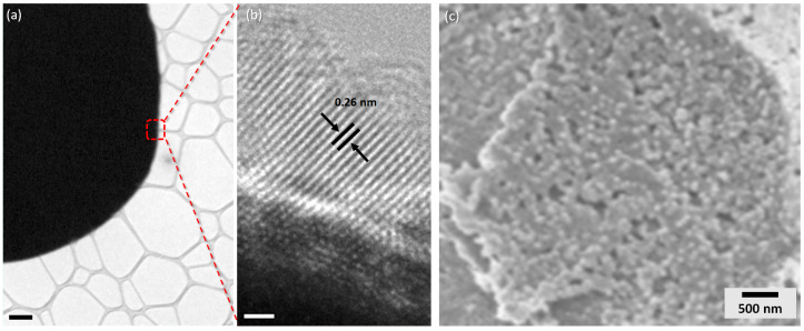
Figure 4.
(a) Transmission electron microscopy (TEM) images of a single ZnO microsphere and (b) high magnification of the edge of the microsphere showing the lattice fringes. (c) High-magnification SEM images of a zinc oxide microsphere shell showing the porous nanosized zinc oxide particles. Scale bars: (a) 1 m; and (b) 1 nm.

