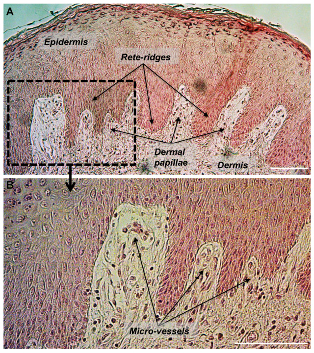
Figure 1.
Histological section of human skin showing the undulated structure of the dermal-epidermal junction with epidermal rete-ridges and dermal papillae. Section of a paraffin-embedded skin biopsy from the abdomen of a 30 years old donor was processed for HES staining. (A) Epidermis, dermis, rete-ridges, and dermal papillae are shown. (B) Micro-vessels are shown within dermal papillae. Scale bars = 100 μm.

