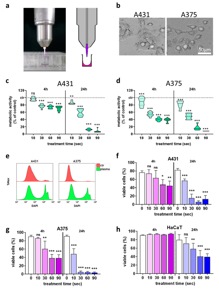
Figure 1.
Plasma treatment inactivated skin cancer cells in a treatment time-dependent fashion. (a) atmospheric pressure argon plasma jet kINPen; (b) representative brightfield image of the two cancer cell types used in this study; (c,d) normalized metabolic activity 4 h and 24 h after plasma treatment in A431 (c) and A375 (d) cells; (e) representative overlay flow cytometry histograms of the terminal cell death dye DAPI in cells with and without plasma treatment at 24 h; (f–h) quantification of viable A431 (f), A375 (g), and HaCaT (h) cells using flow cytometry. Data are the mean of three independent experiments. Statistical analysis was performed using one-way ANOVA (* = p < 0.01, ** = p < 0.01, *** = p < 0.001). ns = not significant, ctr = control.

