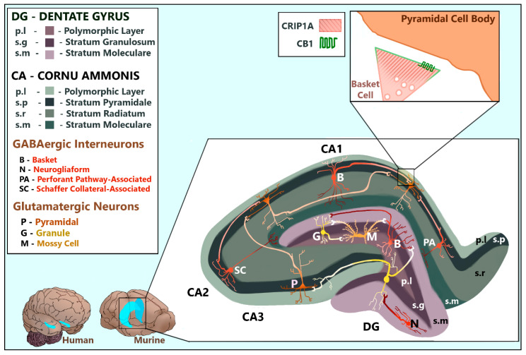
Figure 4.
The mammalian hippocampal formation. CRIP1a is found dispersed in the presynaptic termini of GABAergic interneurons and glutamatergic neurons including DG granule cells, DG hilar mossy cells, and CA pyramidal cells. CB1R is localized to the presynaptic membranes of GABAergic interneurons and glutamatergic neurons including DG hilar mossy cells and CA pyramidal cells. This figure compares the relative size and location of the hippocampal formation in human and murine brains. The enlarged murine hippocampus depicts the shapes, positions, projections, and common synapses of relevant neurons within the layers of the dentate gyrus and hippocampus proper, viewed as a coronal slice. These cellular structures and positions were artistically rendered based upon data reviewed by [36,37,38,39,40]. The interneuron subtypes depicted were chosen based upon those with the strongest evidence of CB1R expression, as reviewed by Pelkey and colleagues [36]. The enlarged synapse depicts the subcellular localization of CB1R and CRIP1a in a CCK+ basket cell interneuron synapsing at a pyramidal soma, notably deficient of both proteins [36]. The synapse conformation was artistically rendered based upon data published by Dudok and colleagues [41].

