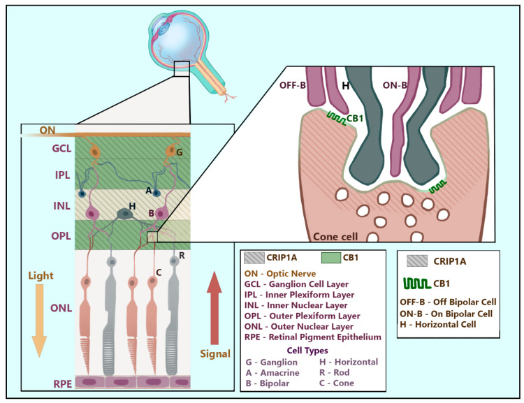
Figure 5.
Distribution of CB1R and CRIP1a in the retina. CRIP1a (shown here as diagonal lines) is found dispersed throughout presynaptic regions of glutamatergic and GABAergic retinal neurons spanning the ganglion cell layer through the outer plexiform layer. CB1R is localized to the presynaptic membranes of retinal synapses in the ganglion cell layer and both inner and outer plexiform layers. This figure artistically renders the positions and common synapses of relevant neurons within the layers of the retina while superimposing identified regions of CRIP1a and CB1R expression. The enlarged synapse depicts the known region of CRIP1a and CB1R co-expression at the presynaptic zone of a cone cell in the outer plexiform layer. This artistic rendering was designed based on the described interaction between cone cells and bipolar cells by Chapot and colleagues [63] and on the shape and location of retinal neuronal cell types described in Neuroscience, 2nd Edition [64].

