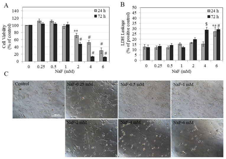
Figure 2.
Viability of human epidermal melanocytes darkly-pigmented (HEMn-DP) cells after 24 and 72 h treatment with NaF assessed by (A) MTS metabolic activity and (B) LDH leakage, ** p < 0.01, $ p < 0.001, and # p < 0.0001 vs. untreated control. One-way ANOVA followed by Dunnett’s post-hoc test was used. (C) Representative images of HEMn-DP cells treated with NaF (0–6 mM) for a period of 72 h, images were taken at 20× magnification. All data are mean ± SEM of at least three independent experiments (n = 3).

