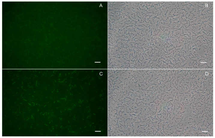
Figure 1.
Fluorescence and non-fluorescence phase contrast microscopy images of A. pleuropneumoniae wild type (wt) (A and B respectively) and pMK_apfas-ACPm-vacJ cells (C and D respectively) after staining with fluorescent CoA 488. Scale bars represent 10 μm. Results presented are representative of three independent experiments.

