Figure 4.
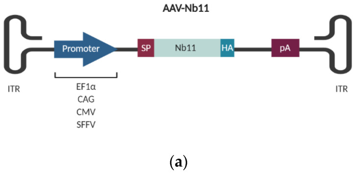
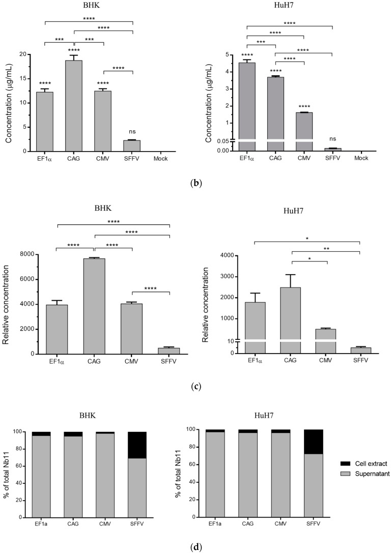
Quantification of Nb11 expressed from BHK and HuH-7 eukaryotic cell lines. Cells were transfected with 2 µg/well of adeno-associated virus (AAV) plasmids coding for Nb11 under the indicated promoters, and 48 h later supernatants and cell extracts were collected for analysis. (a) Schematic representation of AAV vectors coding for Nb11 under the control of four different promoters. (b) Nb11 levels in supernatants of transfected BHK and HuH-7 cells, measured by a hPD-1 binding ELISA. Data represent the mean + SEM. (c) Relative levels of Nb11 in supernatants normalized with Nb11 DNA levels quantified from transfected cells, expressed as Nb11 concentration (ng/mL)/2ΔCt. (d) Analysis of the secretion of Nb11 in transfected BHK and HuH-7 cells. The percentage of secretion was calculated by quantifying the total amount of Nb11 present in supernatants and cell lysates and determining the percentage of Nb11 present in each type of sample. Asterisks above bars indicate comparison of each group with mock. Other comparisons are indicated by horizontal bars. * p < 0.05, ** p < 0.01, *** p <0.001, **** p < 0.0001, ns: not significant. ITR: AAV inverted terminal repeats; EF1α: human elongation factor 1α promoter; CMV: human cytomegalovirus promoter; SFFV: Spleen focus-forming virus promoter; SP: signal peptide; HA: hemagglutinin tag; pA: synthetic polyadenylation sequence.


