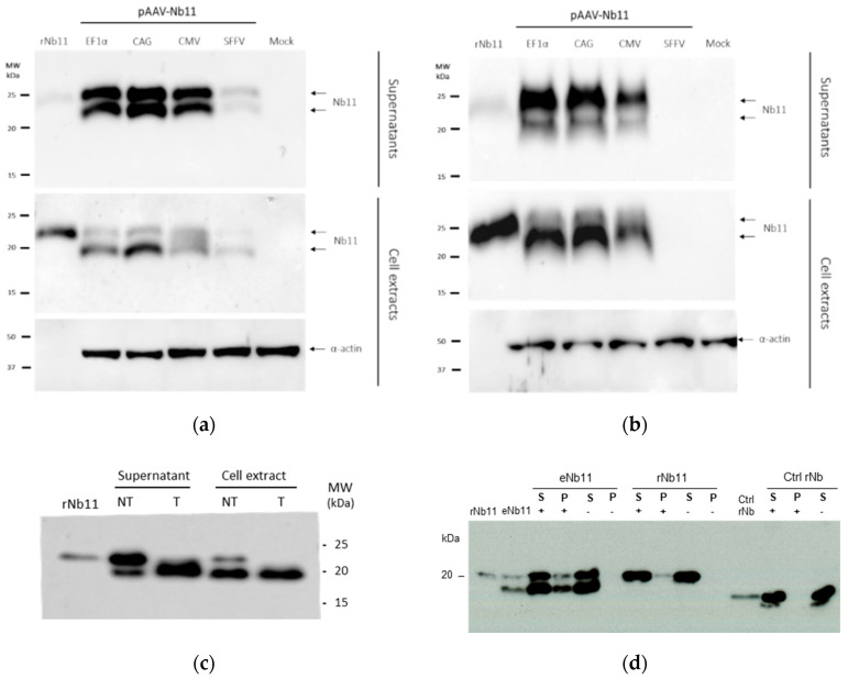
Figure 5.
Western blot analysis of Nb11 expressed from eukaryotic cells In vitro. Supernatant (S) and cell extracts from (a) BHK and (b) HuH-7 cells transfected with AAV plasmids coding for Nb11, under the promoters indicated above the gels, were analyzed by Western blot using an anti-HA antibody. Nb11 in HuH-7 cell samples transfected with pAAV-SFFV-Nb11 could not be detected in this analysis. Twenty nanograms of rNb11 (produced in bacteria) were loaded as control. (c) Analysis of glycosylation in supernatant and cell extracts from BHK cells transfected with pAAV-EF1α-Nb11 that were treated (T) or non-treated (NT) with a mix of N-and O-glycosidases. (d) Immunoprecipitation of Nb11 using mPD-1. Supernatants from BHK cells transfected with pAAV-EF1α-Nb11 (eNb11) or rNb11 were incubated with microbeads coated (+) or uncoated (−) with mPD-1-Fc. After incubation, microbeads were magnetized and the mPD-1-bound fraction (P: precipitated) or unbound fraction (S: soluble) were separated for Western blot analysis using an anti-HA antibody. Ctrl rNb, recombinant control Nb having an HA tag with no relevant specificity.

