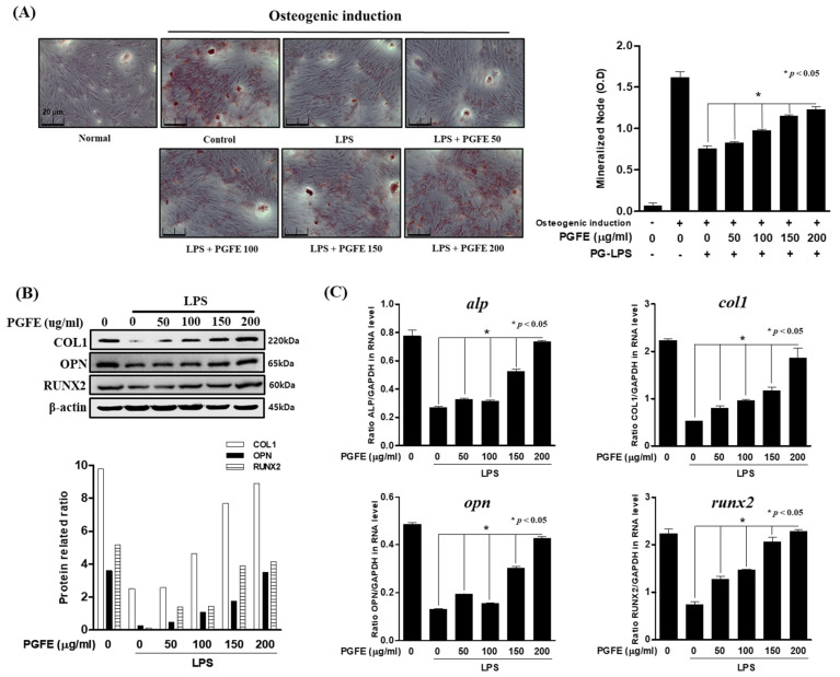
Figure 6.
The effect of PGFE on osteogenic induction in HPDL cells. (A) The periodontal ligament (PDL) cells were pretreated with 50 100, 150 and 200 μg/mL for 6 h and then incubated with LPS for 7 days. (B) The protein expressions were confirmed by Western blot analysis. (C) The mRNA levels of alp, col1, opn, and runx2 were measured by real-time PCR. The results were normalized to gapdh or β-actin expression. * p < 0.05 was considered significant compared to only the PG-LPS treated group. Scale bars denoted 20 μm.

