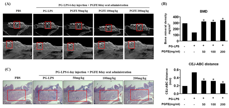
Figure 9.
Inhibitory effect of PGFE on periodontitis in PG-LPS-induced in vivo model. (A) Micro-CT analysis of newly formed bone by PGFE, the red mark is the PG-LPS injection area. (B) Quantification of distance between CEJ and ABC, and bone mineral density. Analysis tables were determined using CTAn software. (C) Histological analysis of the periodontium using H&E staining. * p < 0.05 vs. only PG-LPS treated group. Scale bars denoted 20 μm.

