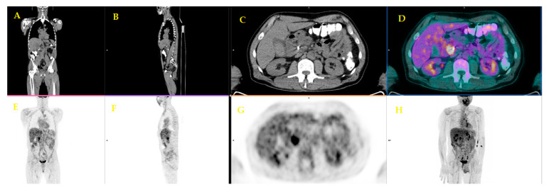
Figure 4.
A fifty-six-year-old patient underwent staging FDG PET/CT with positive (ce)CT examination for local pancreatic lesion and equivocal for distant metastases. FDG PET/CT revealed a focal uptake on the head of the pancreas (C,D,G). (Images from Nuclear Medicine Unit, Padova University Hospital). Legend: A = Coronal image of low-dose CT; B = Sagittal image of low-dose CT; C = Axial image of low-dose CT; D = PET/CT fused image; E = Coronal image of PET; F = Sagittal image of PET; G = Axial image of PET; H = Maximum intensity Projection (MIP).

