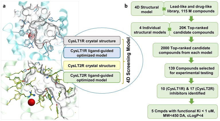
Figure 1.
Overview of the prospective VLS for CysLTRs. (a) Optimized ligand pockets for CysLT1R (blue) and CysLT2R (green) and the composition of the 4D Screening model. Key water in the optimized pocket of CysLT1R and CysLT2R is shown by red sphere. (b) Flowchart of screening and ligand identification procedure.

