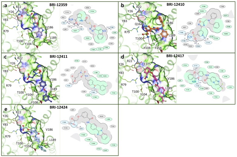
Figure 6.
Predicted binding posesof the experimental hits in the refined CysLT1R binding pocket. Ligands are shown is stick presentation. Receptor is shown in cartoon and thin stick presentation with carbons colored green. The pocket is shown as transparent surface colored according to properties (green: hydrophobic, blue: H-bond donor, red: H-bond acceptor). 2D diagrams depict ligand interactions with receptor residues, with H-bonds shown as dashed lines and distances in Å.

