Figure 5.
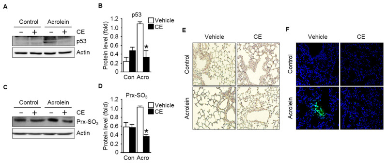
Effect of CE on p53 activation in pulmonary tissues exposed to acrolein: (A) p53 level in the lung sections was determined by immunoblot analysis. Actin served as an internal control; (B) quantification of p53 levels normalized to the expression of actin is shown; (C) immunoblot analysis of Prx-SO3 level, a marker for intracellular reactive oxygen species (ROS) formation, in acrolein-exposed lung tissues with or without pre-treatment with CE. Actin was used as the loading control; (D) a graph depicting the quantification of the relative abundance of the Prx-SO3 protein levels is shown. Each protein sample was subjected to 8–12% SDS-PAGE and immunoblotted with related antibodies as described in the text; (E) representative histochemical staining of the lung tissues (20× magnification) with 3,3′-diaminobenzidine (DAB) for the measurement of the level of intracellular H2O2; (F) representative images of immunohistochemical analyses of lipid peroxidation adducts, 4-hydroxynonenal (4-HNE) in the lung tissues (400× magnification). Nuclei were counterstained with 4′,6-diamino-2-phenylindole (DAPI). Results are expressed as the mean ± S.D. (n = 3–6 mice per group). p < 0.05 was considered statistically significant. * indicates statistical significance compared to the acrolein-treated group. Con, control; Acro, acrolein.

