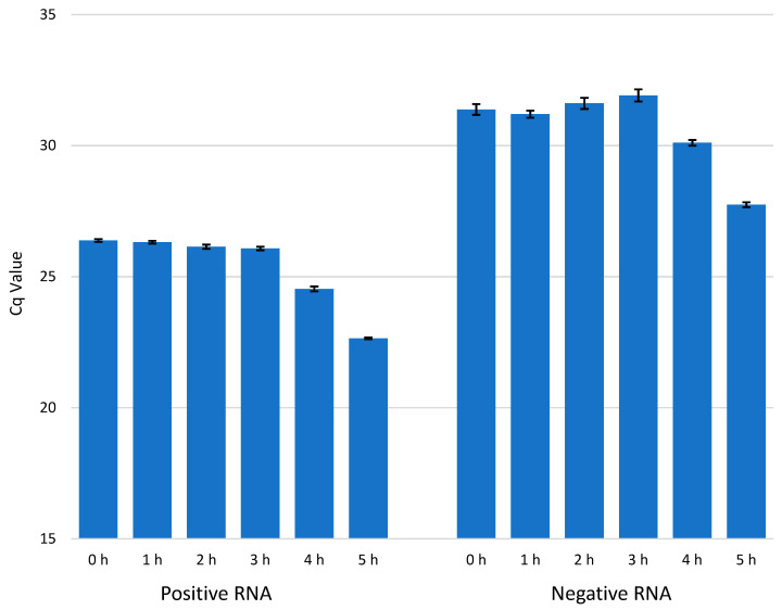
Figure 4.
The amount of viral +RNA and −RNA in CVA9-infected cells at timepoints 0–5 h p.i. The amount of +RNA at timepoints 0–5 h p.i. is shown on the left, and the amount of −RNA at timepoints 0–5 h p.i. is shown on the right. The bars are averages calculated from triplicate samples, and the error bars are standard deviations from the average values.

