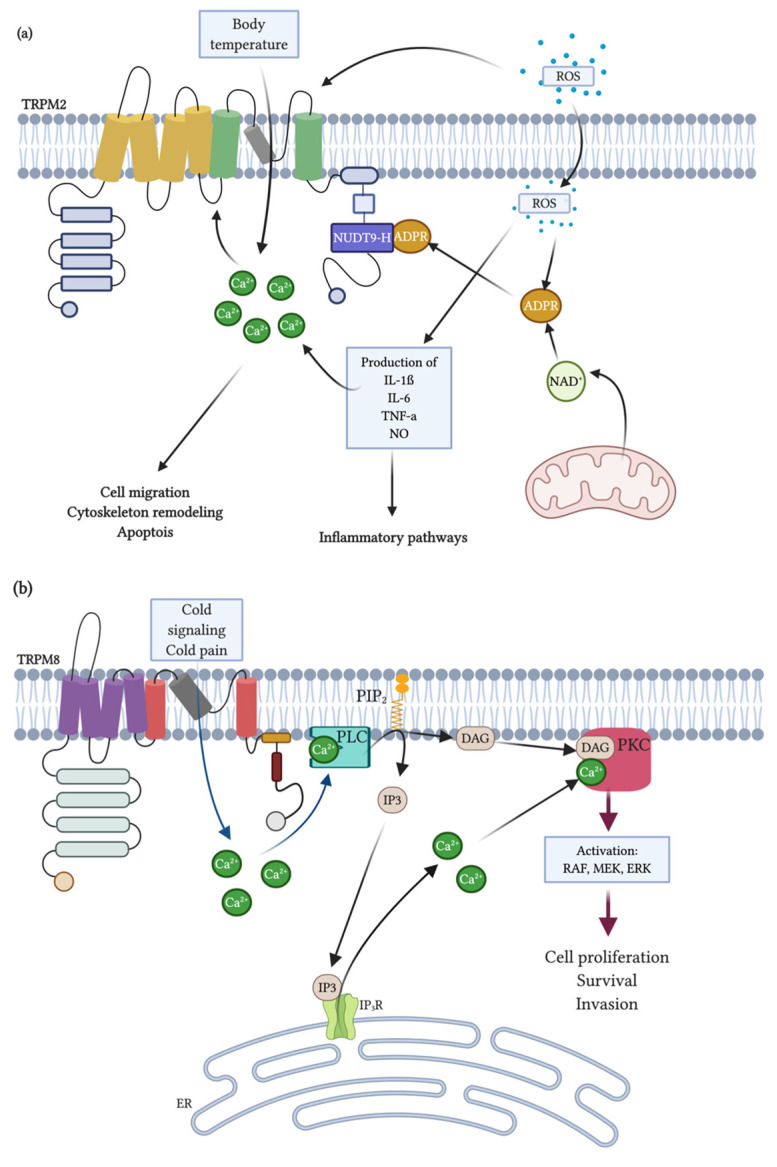
Figure 3.
Signaling mechanisms for TRPM2 and TRPM8 activation. (a) NAD+ and reactive oxygen species (ROS), including H2O2, accumulate during inflammation and tissue damage may trigger TRPM2. NAD+ may be converted to ADPR and cADPR. ROS can also cross the plasma membrane and mobilize ADPR from mitochondria and both ROS and cADPR can synergize with ADPR to activate TRPM2. Additionally, ADPR is generated from NAD+ via poly-ADPR during ROS-induced damage through the activation of PARP (poly(ADP-ribose) polymerase)-ADPR-dependent mechanisms in inflammatory cells. Free cytosolic ADPR can act on the NUDT9-H of TRPM2 channels, enabling Ca2+ influx across the plasma membrane and/or release of intracellular Ca2+, raising the Ca2+ concentration in the cytosol. On the other hand, ROS induces cytokine production (pro-inflammatory response), which may alter intracellular calcium levels. A Ca2+ increase will activate different physiological processes including gene expression through Ca2+-dependent signaling pathways such as apoptosis, cell migration and cytoskeleton remodeling. Moreover, TRPM2 may detect increased temperatures to prevent overheating, limiting the fever response. (b) Cold temperature, menthol or icilin can stimulate the activity of the TRPM8 ion channels. Upon activation, the TRPM8 channel changes conformation and permits extracellular Ca2+ to flow through it across membranes. This results in the activation of Ca2+-sensitive PLC and the hydrolysis of PIP2, which produces inositol 1,4,5-triphosphate (IP3) that causes the release of Ca2+ from the intracellular stores and generates diacylglycerol (DAG). The elevated Ca2+ level or DAG can activate protein kinase C (PKC), which, in turn, activates RAF in the ERK pathway. Therefore, transcriptional activation of genes stimulates cellular proliferation, survival and invasion, which contribute to cancer growth and metastasis. Created with BioRender.com.

