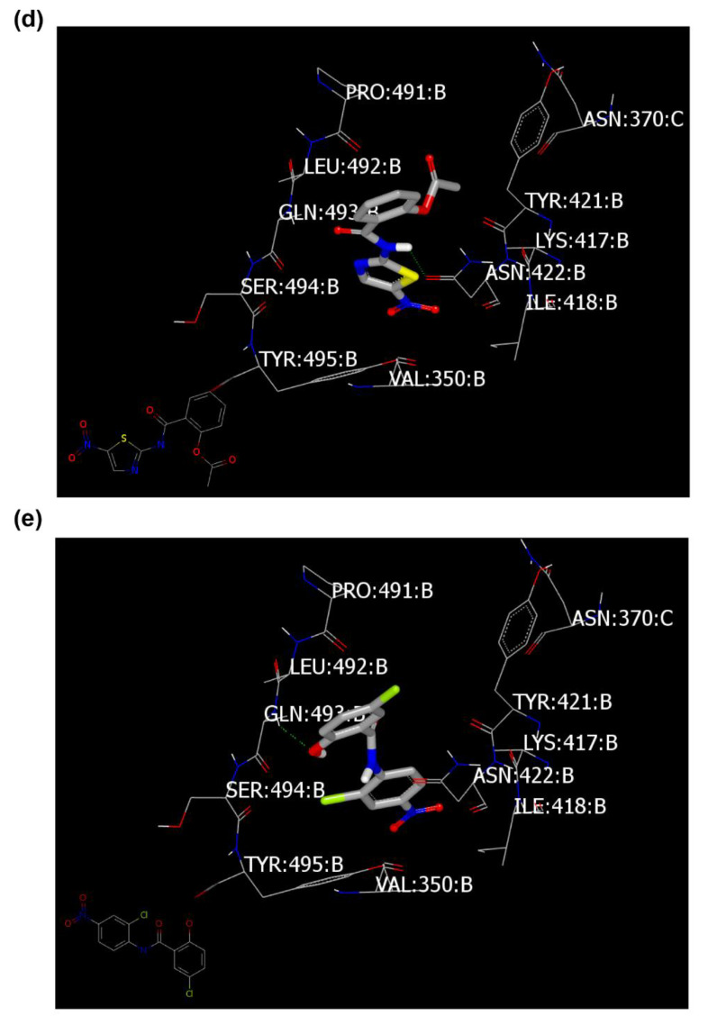
Figure 4.
Visual representation by VIDA for docking with spike glycoprotein (PDB ID: 6vsb). (a) Standard ligand docked inside the receptor (HB in green color), (b) ligand inside the inner grid for validation, (c) Azithromycin docked peripherally, (d) Nitazoxanide docked with formation of weak HB (green color), and (e) Niclosamide docked with formation of strong HB (green color).


