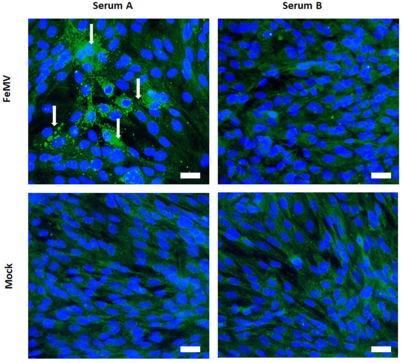
Figure 3.
Serology of analyzed guigna serum samples from Chile. Representative result of a serological analysis via immunofluorescence assay. FeMV-specific antibodies were detected in a guigna serum sample (serum A) illustrated by fluorescence staining of perinuclear and cytoplasmic viral inclusion bodies (white arrows). In comparison, an IFA-negative sample (serum B) is shown on the right. Scale bar represents 20 µm.

