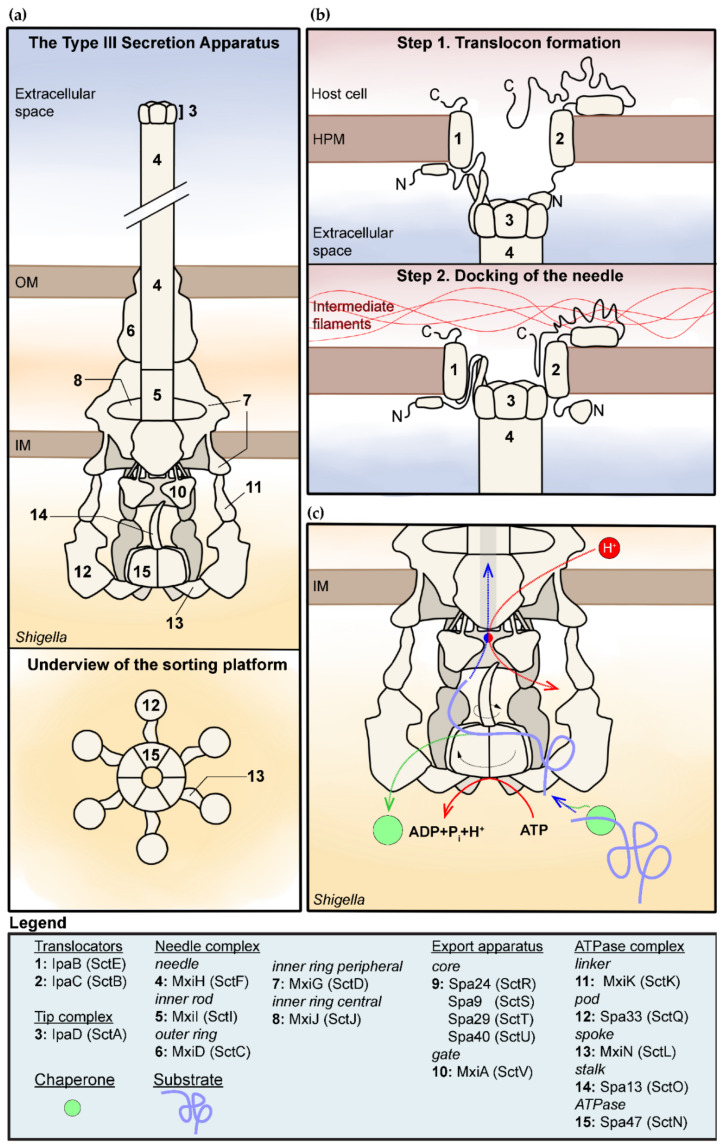
Figure 1.
Structure and function of the type III secretion apparatus (T3SA) in Shigella spp. (a) Overview of the structure of the inactive T3SA. Note that the tip complex and cytosolic components of the T3SA are represented with a 3D perspective while the body is represented as a flat longitudinal cross section [16,17,31,32]. The bottom panel represents the sorting platform viewed from the cytosol of the bacterium. (i.e., viewed from the underside). (b) Model for the formation of the translocon and mutual interaction with the tip complex, the host plasma membrane, and the intermediate filaments [33,34]. (c) This is a model for the secretion of T3SA substrates summarizing elements discussed in the text. It indicates the role of the chaperone and ATPase–stalk complex in the unfolding of the substrates (purple) through rotation induced by ATP hydrolysis [35,36], and of the gate of the export apparatus in the creation of a proton motive force required for substrates secretion [37,38]. Movement is represented by dashed arrows; rotation is represented by curved arrows. The coupling of the protein substrates–proton antiporter is represented by the red–blue circle. The legend indicates the name of the various components in Shigella and in the unified nomenclature in parentheses.

