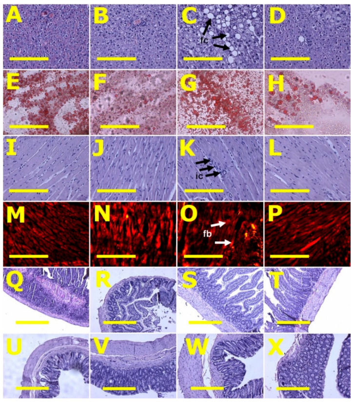
Figure 2.
Liver fat structure using haematoxylin and eosin stain (A–D) and oil red O stain (E–H); left ventricular structure—heart inflammation (I–L) using haematoxylin and eosin stain and heart fibrosis (M–P) for collagen using picrosirius red staining; ileum (Q–T) and colon (U–X) structure using haematoxylin and eosin stain in corn starch diet-fed rats (A,E,I,M,Q,U), corn starch diet-fed rats supplemented with Caulerpa lentillifera (B,F,J,N,R,V), high-carbohydrate, high-fat diet-fed rats (C,G,K,O,S,W) and high-carbohydrate, high-fat diet-fed rats supplemented with Caulerpa lentillifera (D,H,L,P,T,X). Fat cells = fc; inflammatory cells = ic; fibrosis = fb. Scale bar is 200 μm for (A–P) (20×) and 100 μm for (Q–X) (10×).

