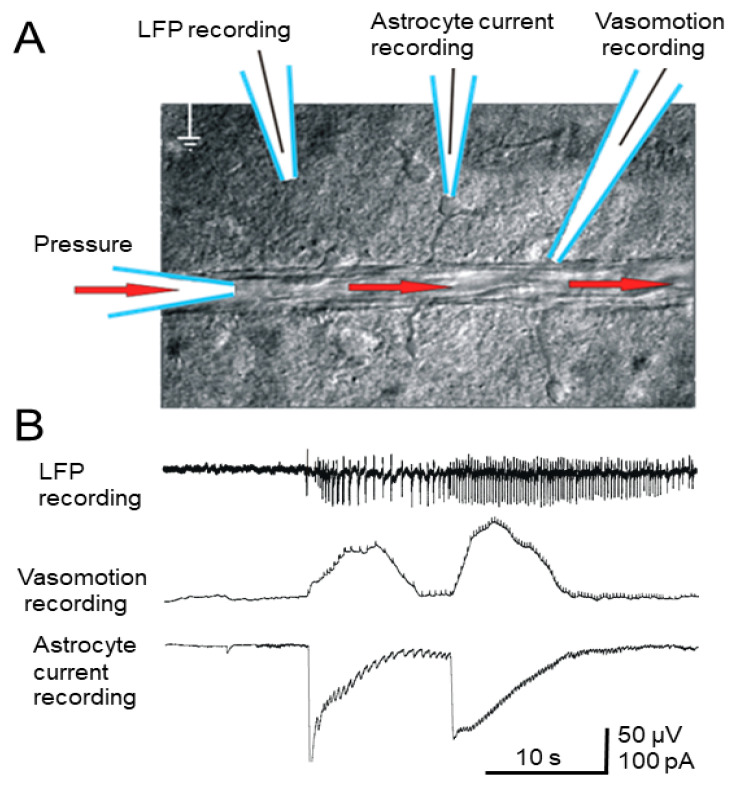
Figure 4.
(A) Positioning of the blood vessel perfused with ACSF (30 torr) and recording electrodes. (B) Simultaneous extracellular LFP, vasomotor, and astrocyte patch-clamp recording after 4-AP application (see text). Low-voltage, high-frequency components superimposed on the vasomotor response, but did not reflect actual vasodilation, reflecting neural spikes.

