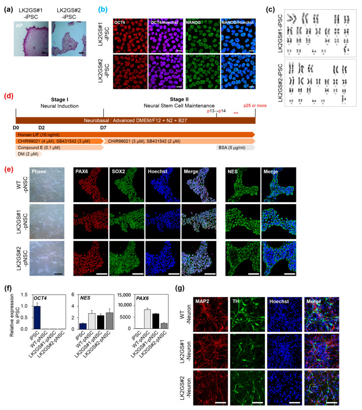
Figure 1.
Characterization of pNSCs differentiated from PD patient-specific induced pluripotent stem cells (iPSC). (a) Alkaline phosphatase (AP) activity of established iPSC (Scale bar, 200 μm). (b) Immunostaining of established iPSC for human pluripotent stem cell markers, OCT4, and NANOG (scale bar, 50 μm). Counterstain, Hoechst33342. (c) Karyotypes of established iPSC. (d) A schematic diagram of the differentiation protocol used to obtain pNSC from iPSC. *** p < 0.001. (e) Representative morphology and immunostaining of pNSC with neural stem/progenitor markers, PAX6, SOX2, and NES (scale bar, 100 μm). (f) mRNA expression levels of human pluripotent stem cell marker (OCT4) and neural stem cell markers (NES and PAX6) in differentiated pNSCs. (g) Representative fluorescence image of differentiated neurons with neuronal cell marker, MAP2, and dopaminergic neuron marker, TH (scale bar 100 μm). LK2GS; PD patient-derived cells harboring the Gly2019Ser mutations in the leucine-rich repeat kinase 2 gene, WT; healthy control cells.

