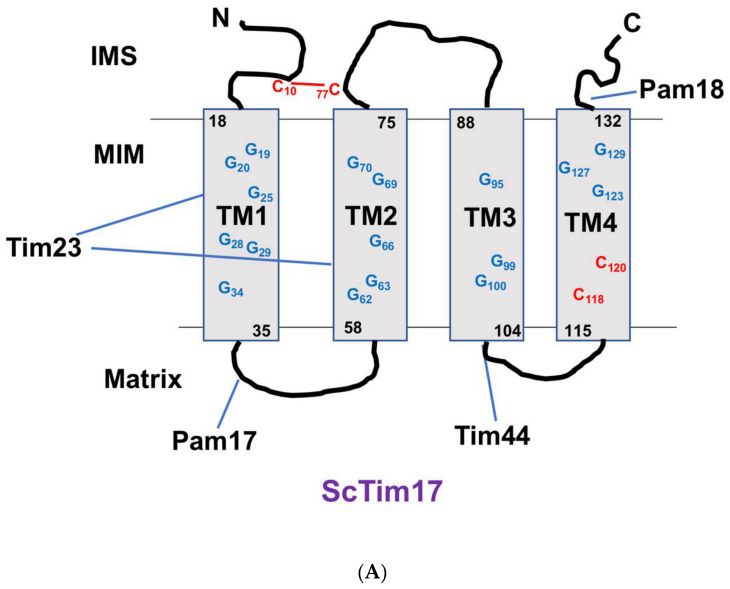
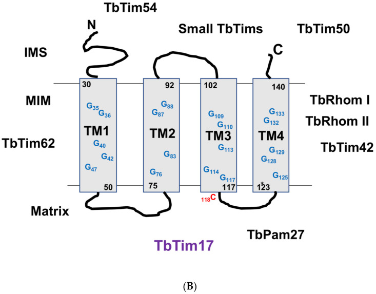
Figure 2.
Schematics of the secondary structures of Tim17 of the Saccharomyces cerevisiae (A) and Trypanosoma brucei (B). Four transmembrane domains (TM1–TM4) and the hydrophilic loop regions are shown. Glycine (G in blue) residues within the TMs and cysteine (C in red) residues are indicated. Numbers indicate the position of these Gs and Cs in ScTim17 and TbTim17 sequences. Cs that form the disulfide bond in ScTim17 are connected with lines. IMS, intermembrane space; MIM, mitochondrial inner membrane; and the matrix are indicated. Interacting regions of ScTim17 with Tim23, Pam17, Pam18, and Tim44 are indicated. Interacting regions of TbTim17 with other subunits are yet to be determined. However, the known localization of these subunits in the IMS, MIM, and matrix is shown.


