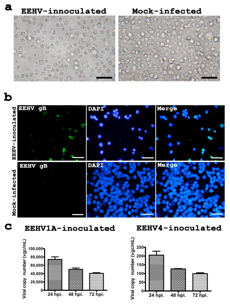
Figure 2.
Cell morphology, immunolabeling for EEHV gB and determination of EEHV terminase genes in the U937 cells after inoculation with EEHV. At 72 hpi, although no cytological changes were observed in the U937 cells (a), immunolabeling for the EEHV gB was shown to be positive by immunofluorescence in the EEHV-inoculated group (b). Quantitative PCR presented as viral genome copies (vgc/mL) of the U937 cell culture supernatant at 24, 48 and 72 hpi indicated that there was a reduction of EEHV in both the EEHV1A-inoculated and EEHV4-inoculated cells (c). Scale bars in (a) ~200 µm, in (b) ~300 µm.

