Figure 3.
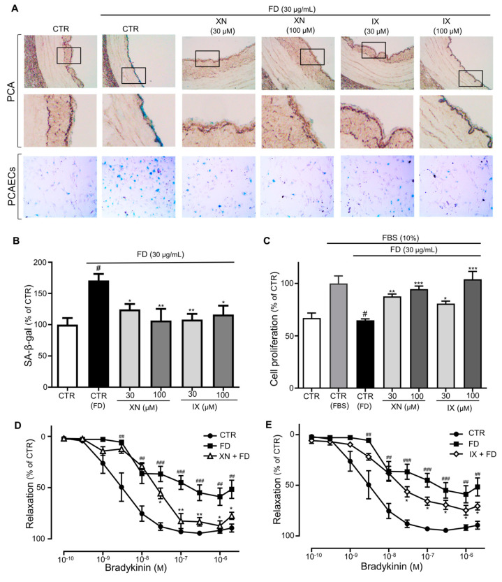
XN and IX prevent FD-induced premature senescence. (A) PCA rings and PCAECs were incubated in FD alone or FD in combination with XN or IX (30, 100 μg/mL) before determination of SA-β-gal activity by X gal staining. Representative images of SA-β-gal-stained PCA rings and PCAECs. (B) Cumulative data of SA-β-gal activity as a percentage of the control. Data are expressed as the mean ± SEM (n = 6); # p < 0.05 vs. CTR; * p < 0.05, ** p < 0.01 vs. FD alone (CTR-FD). (C) Dose-dependent increases in cell proliferation after FD and XN/IX (10,30 μM) treatment. Data are expressed as the mean ± SEM (n = 6); # p < 0.05 vs. FBS alone (CTR-FBS); * p < 0.05, ** p < 0.01, *** p < 0.001 vs. FD alone (CTR-FD). (D,E) Concentration–relaxation curves of FD-exposed aortic rings treated with XN and IX (100 μM) in response to bradykinin. Data are expressed as the mean ± SEM (n = 7–10); ## p < 0.01, ### p < 0.001 vs. CTR; * p < 0.05, ** p < 0.01 vs. FD alone (FD).

