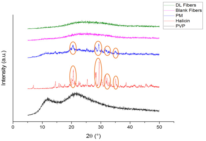
Figure 4.
X-ray diffraction (XRD) patterns of PVP, halicin, PM, blank and drug-loaded fibers showing that the drug is in the crystalline form (presence of characteristic peaks) while the polymer is in the amorphous form (broad halos). The presence of halicin distinct peaks is also present in the PM which are absent in the drug-loaded fibers indicating the molecular dispersion of the drug within the fibers.

