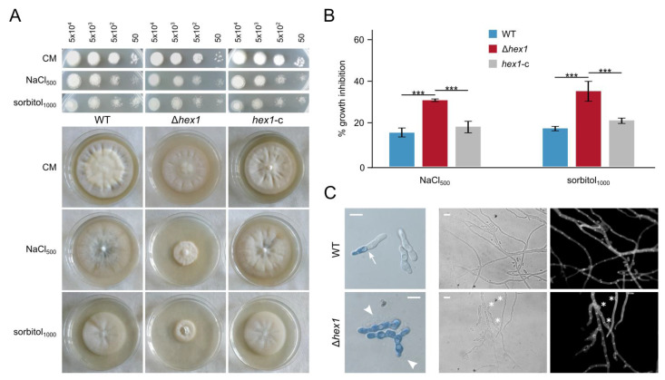
Figure 4.
Effects of hyperosmotic stress on Δhex1. (A) Responses of the wild-type, the deletion (Δhex1), and the complemented (hex1-c) strains to NaCl and sorbitol, with regard to germination (top; the number of inoculated conidia per spot are provided above the images; growth for 3 days) and radial growth (bottom; growth for 18 days). CM: CzD-CM. (B) Relative growth inhibition by NaCl and sorbitol (each condition was tested in triplicate; bars = SD; statistical testing by one-way ANOVA followed by Tukey’s post hoc test (*** p ≤ 0.001). All concentrations are given in mM. (C) Staining of wild-type and Δhex1 germlings with methylene blue, after growth for 16 h in a hyperosmotic medium of 0.5 M NaCl (left). Arrow: septum. Arrowheads: cytoplasmic bleeding. Morphology of corresponding hyphae under the same conditions. Asterisks: “bubble”-like cells. Cell wall staining using calcofluor white M2R. Bar = 10 μm.

