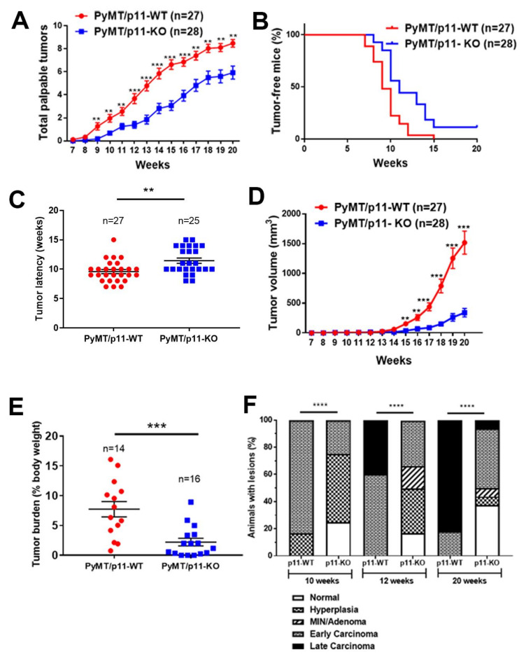
Figure 2.
Tumor initiation, growth, burden, and progression are decreased and delayed in PyMT/p11-KO mice. (A) Appearance of palpable tumors was monitored weekly. (B) Percent tumor-free mice was calculated using Kaplan–Meir analysis (hazard ratio (HR)—2.24, p = 0.0004). (C) Tumor latency was plotted for each mouse based on the first appearance of palpable tumor. (D) Tumor volume was measured weekly using calipers. Total volume was plotted to represent tumor growth rate. (E) Total tumor weight at endpoint (20 weeks) was determined as a percentage of body weight. Data are a compilation of three independent experiments, except (D), which shows data from one independent experiment. Significance was determined by non-parametric t-test (Mann Whitney U test–unpaired, non-parametric test for means). (F) Mammary glands and tumors from PyMT/p11-WT and PyMT/p11-KO mice were harvested at 20 weeks, formalin-fixed, embedded, and sectioned at 5 µm thickness. The H&E and smooth muscle α-actin-stained sections were classified into histopathological stages by a pathologist in a blinded manner. (n = 6 to 12 mice in each group). Statistical analysis was performed by χ2-test (p < 0.0001). The ssterix on each plot represents p-values as follows. ** p < 0.01, *** p < 0.001, **** p < 0.0001.

