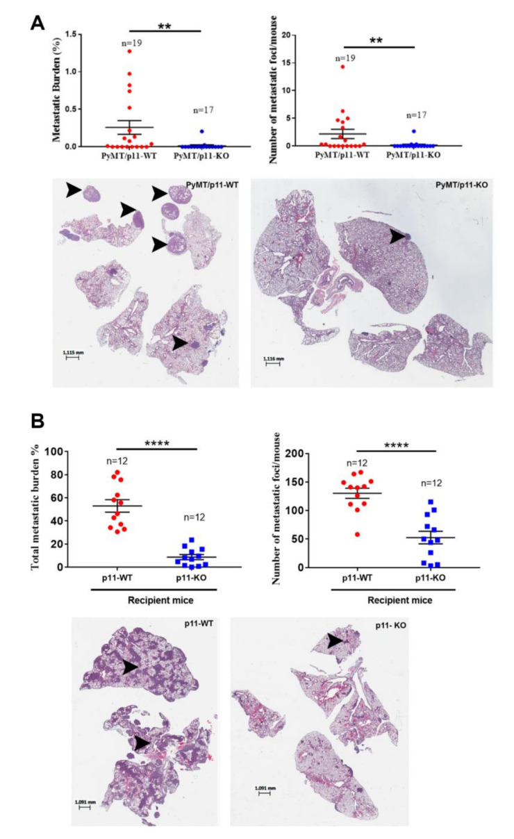
Figure 4.
Metastasis is diminished in PyMT/p11-KO mice. (A) We evaluated pulmonary metastasis at the 20-week end point in (spontaneous model) PyMT/p11-WT and PyMT/p11-KO mice by microscopic examination of formalin-fixed, H&E-stained lung sections (5 µm). Three lung sections each 100 µm apart were used for staining. Quantification was performed using Aperio image analysis software (Imagescope). Mean values for three sections were used to calculate the metastatic burden and foci values. (A) We evaluated the metastatic burden by measuring the total metastatic area and total lung area, followed by normalization (A—upper left panel). Mann Whitney U test showed statistical significance with p value of 0.0062. The number of metastatic foci per mouse lung section was determined by manual counting of images (A—upper right panel). Mann Whitney U test showed p value of 0.0069. Lower panels: Representative lung images from WT and KO mice. (B) Experimental metastasis assay. We injected 2.5 × 10⁵ Py8119 (p11-WT levels) cells into p11-WT and p11-KO mice (n = 12 mice per group). The lungs were harvested after 14 days, formalin-fixed, and sectioned at 5 µm as described above. Mann Whitney non-parametric t-test (for means) for statistical significance was performed. Metastasis was pooled and combined from two independent experiments (n = 6 each). p < 0.0001. Lower panels: representative lung images from WT and KO mice. The asterix on each plot represents p-values as follows. ** p < 0.01, **** p < 0.0001.

