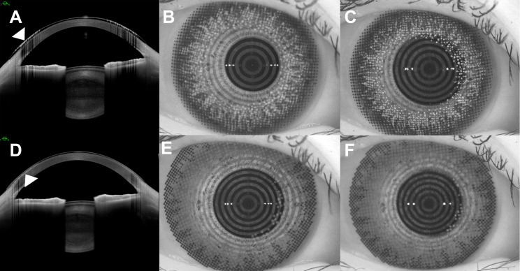
Figure 1.
Arrow indicates the location of the pigment in the SCL front or back surface by anterior segment optical coherence tomography (A and D), and photos of ring mire by the keratograph (B, C, E and F). (B) When we focus on the ring mire, pigment located at the front surface of the SCL blurs. (C) When we focus on the pigment, the ring mire blurs. (E) When we focus on the pigment located at the back surface of the SCL, the ring mire is also clear. (F) We do not focus on the lens surface of SCL because of absence of target.
Abbreviation: SCL, soft contact lens.

