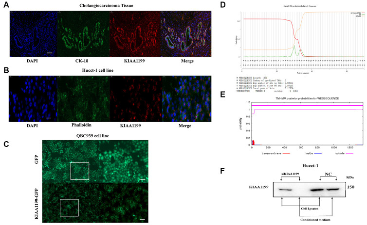
Figure 2.
(A) Immunofluorescence localization of KIAA1199 and CK-18 in cholangiocarcinoma tissue (red, KIAA1199; green, CK-18; blue, DAPI). (B) Immunofluorescence localization of KIAA1199 and Phalloidin in Hucct-1 cell line (red, KIAA1199; green, Phalloidin; blue, DAPI). (C) Microscopic determination of KIAA1199 cellular localization using QBC939 cells transfected with green fluorescent protein (GFP), KIAA1199-GFP chimeric cDNAs. (D) KIAA1199 had signal peptides. (E) KIAA1199 has 602 amino acids, all of which are extracellular, and there is no transmembrane domain (TMD). (F) Western blot analysis was performed to detect KIAA1199 in cell lysates and conditioned culturing medium from Hucct-1 and Hucct-1 knockdown KIAA1199 (siKIAA1199).

