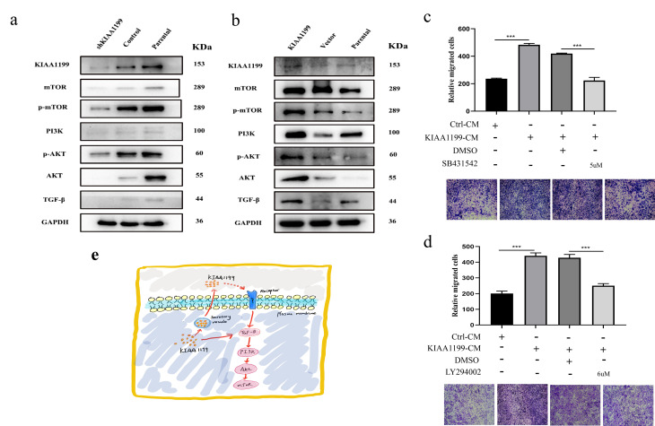
Figure 8.
The expression of KIAA1199 and TGF-β-PI3K-Akt pathway-associated proteins by western blot analyses. (A) Western blot analysis of KIAA1199 and TGF-β-PI3K-Akt pathway-associated proteins in KIAA1199 silenced Hucct1 cell line. (B) Western blot analysis of KIAA1199 and TGF-β-PI3K-Akt pathway-associated proteins in KIAA1199 overexpressed QBC939 cell line. (C) QBC939 were pretreated with TGF-β inhibitor (SB431542, 5 μM) for 2 h and transwell migration assay was performed in the absence or presence of KIAA1199 conditioned medium (CM). (D) QBC939 were pretreated with PI3K inhibitor (LY294002, 6μM) for 2 h and trans-well migration assay was performed in the absence or presence of KIAA1199 conditioned medium (CM). (E) KIAA1199-mediated EMT may occur through a non-Smad pathway. At least three independent experiments were preformed, data presented as mean ± SD, *, ** and *** represented P<0.05, P= 0.01 and 0.001 by Student's t-test, between the indicated group.

