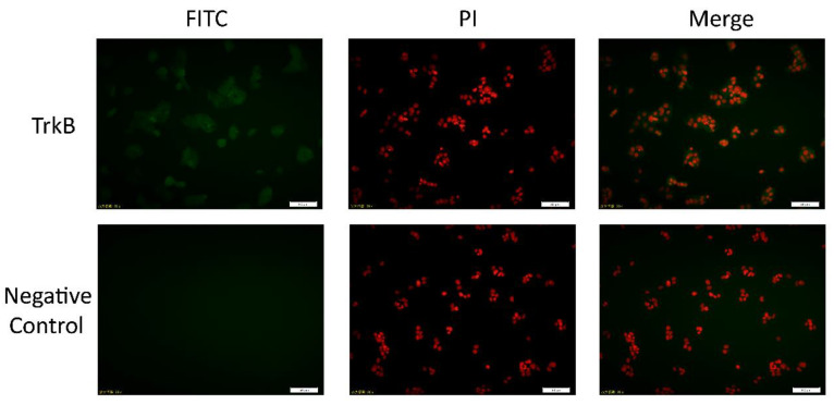
Figure 1.
Determination of TrkB protein in Ishikawa cells by cell immunofluorescence. The Ishikawa cells were incubated with anti-TrkB or primary antibody dilution (negative control), then coupled with FITC-conjugated secondary antibody (FITC; green) and propidium iodide (PI; red) for the detection of nuclei. The picture (merge) was the result of the merging between the two fluorescences. Scale bar = 50 µm. TrkB: tyrosine kinase receptor β.

