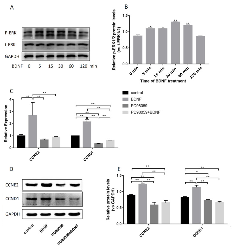
Figure 6.
Effects of BDNF on proliferation of Ishikawa cells through TrkB–ERK1/2 signal transduction pathway. Ishikawa cells were monocultured with 10 μM/mL PD98059 for 30 min and then cocultured with 200 ng/mL BDNF for 24 h. (A) Ishikawa cells were treated with 200 ng/mL BDNF for 0, 5, 15, 30, 60, and 120 min. Protein levels of t-ERK and P-ERK were detected by Western blot analysis. (B) Relative protein levels were analyzed by grey scanning. (C) Relative mRNA expression levels of CCND1 and CCNE2 genes were detected by qRT-PCR. (D) Protein levels of CCND1 and CCNE2 genes were detected by Western blot analysis. (E) Relative protein levels of CCND1 and CCNE2 genes were analyzed by grey scanning. The expression levels of mRNA and proteins were normalized by the GADPH gene, which plays an important role as an internal reference calibration for qRT-PCR and Western blot analysis. Statistical analysis is shown. * p < 0.05, ** p < 0.01.

