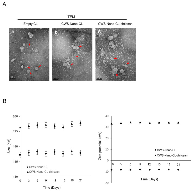
Figure 1.
Characterization of the CWS-loaded formulations. (A) Transmission electron microscopy (TEM) images of empty CL, CWS-Nano-CL, and CWS-Nano-CL-chitosan. The red arrows indicate the prepared liposomes. All of the scale bars indicate 200 nm. (B) Colloidal stability of the CWS-Nano-CL, and CWS-Nano-CL-chitosan. Data are mean ± SD (n = 3).

